You are here
💬 Think about it
Hold your phone far away and the tiny letters blur into one blob. Bring it close and you can read again. Your eyes have a limit. Cells are far smaller than that limit. So how do scientists ever see them? Let us find out.
![2.1 How to Study Cells? - Cell — The Building Bl - [Teachoo] - Concepts](https://cdn.teachoo.com/24e07b90-a187-4b1f-98b5-bb0ad9c60e8c/image-a-size-scale-chart-from-1-kilometre-down-to-0.1-nanometre-showing-where-a-rocket-a-tree-a-human-a-chicken-egg-plant-and-animal-c.webp)
Fig. 2.1 as a Scale — What Each Tool Can See
| Size | Example object | Seen with |
|---|---|---|
| 1 km – 1 m | Rocket, Neem tree, human height, Great Indian Bustard | Unaided eye |
| 0.1 m – 1 cm | Chicken egg | Unaided eye |
| 1 mm | Fish egg, Amoeba | Unaided eye / light microscope |
| 100 µm – 10 µm | Most plant and animal cells, nucleus | Light microscope |
| 1 µm | Most bacteria, mitochondrion | Light microscope |
| 100 nm – 10 nm | Smallest bacteria, viruses, ribosomes | Electron microscope |
| 1 nm – 0.1 nm | Proteins, lipids, small molecules, atoms | Electron microscope |
What is the limit of resolution of the human eye?
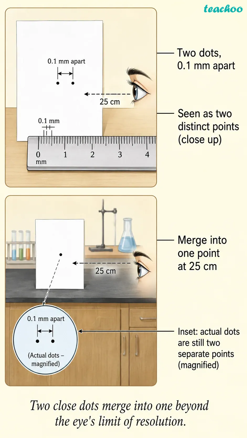
Limit of Resolution = 0.1 mm
From about 25 cm, the eye can separate two points only if they are at least 0.1 mm apart; closer than that they look like one point.
Example:
Two tiny dots drawn very close merge into one when seen from far.
- Resolution means seeing two close points as separate.
- From about 25 cm, our eye separates points 0.1 mm apart.
- Closer than that, they look like a single point.
- So the limit of resolution of our eye is 0.1 mm.
Example:
Two tiny dots drawn very close on paper merge into one dot when seen from far.
Why can't we see a cell with our naked eye?
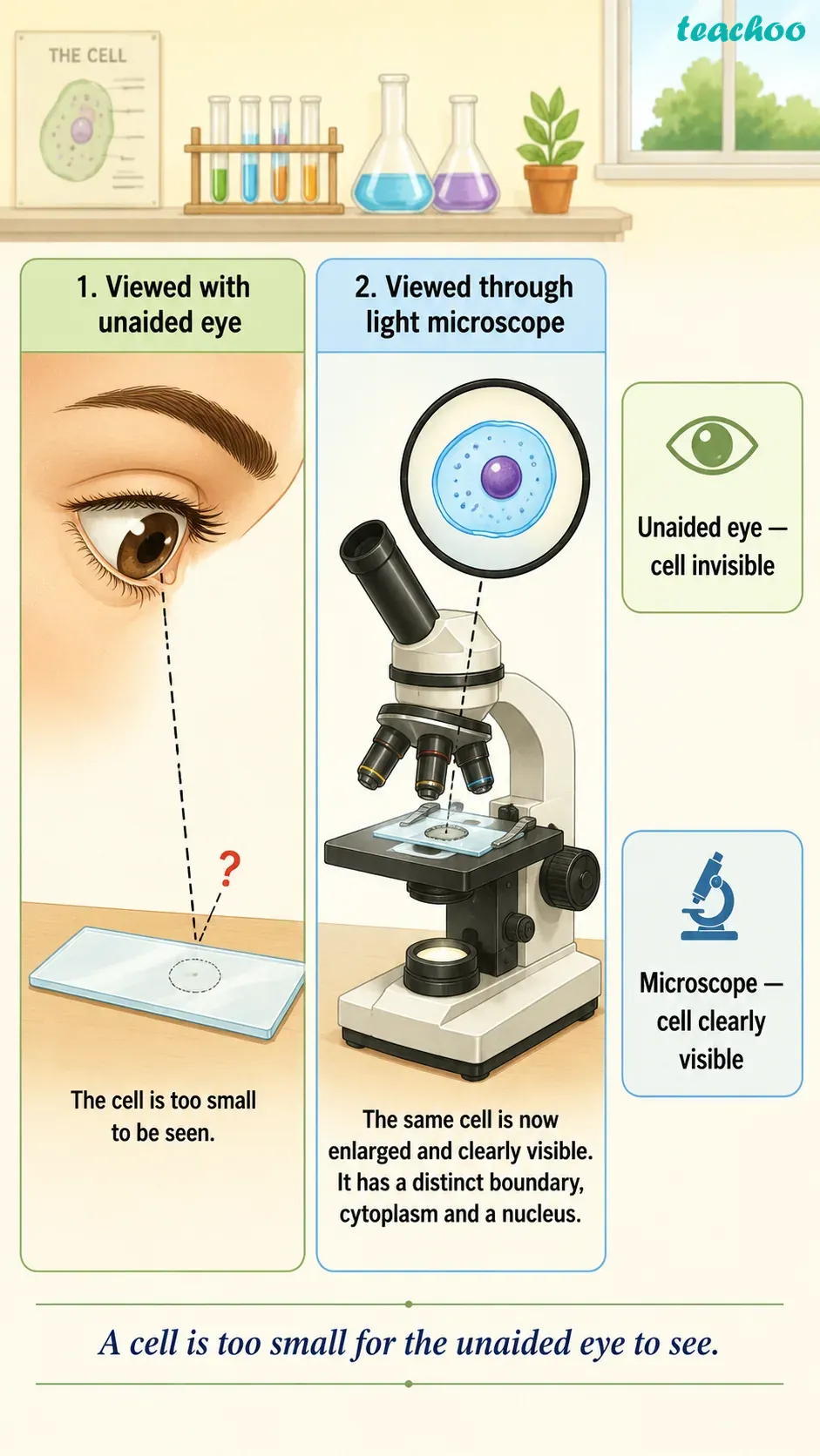
- A cell is usually much smaller than 0.1 mm.
- So it is below the limit of our eye.
- We need tools that make small things look bigger.
How does a microscope help us see tiny things?
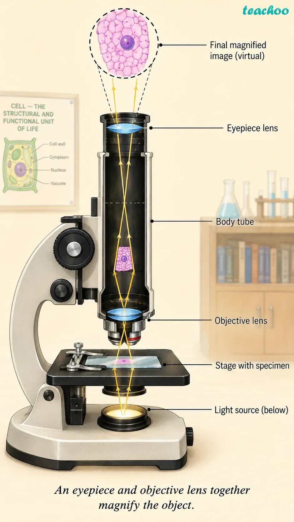
- A convex lens, or a set of lenses, magnifies an object.
- Magnification makes a small object appear larger.
- A microscope uses an objective lens and an eyepiece together.
Example:
A magnifying glass is a single convex lens that makes print look bigger.
Who first saw and named cells?
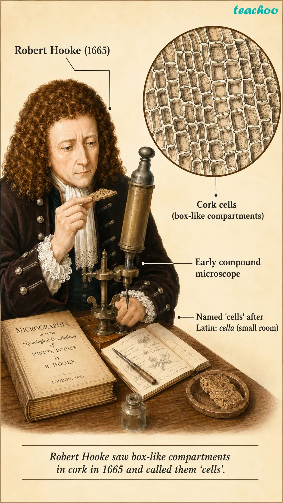
Robert Hooke (1665)
He used a self-designed microscope and saw box-like compartments in a thin slice of cork. He named these boxes 'cells'.
- Robert Hooke was the first to observe a cell in 1665.
- He used a self-designed microscope (about 200–300X).
- He saw box-like compartments in a thin slice of cork.
- He named these boxes "cells".
What are the main parts of a light microscope?
Parts of a Light Microscope
Eyepiece
The lens you look through at the top.
↓
Body Tube
Connects the eyepiece to the objective lens.
↓
Objective Lens
The lens near the object — e.g. 10x, 40x.
↓
Coarse & Fine Knobs
Move the lens up and down to focus the image.
↓
Stage
Flat platform that holds the glass slide.
↓
Mirror / Light Source
Sends light through the slide and lens.
| Part | Job |
|---|---|
| Eyepiece | The lens you look through at the top. |
| Body tube | Connects the eyepiece to the objective lens. |
| Objective lens | The lens near the object (e.g. 10X, 40X). |
| Coarse & fine knobs | Move the lens to focus the image. |
| Stage | The flat platform that holds the slide. |
| Mirror | Reflects light up through the object. |
📷 Image placeholder — upload a photo of a light microscope
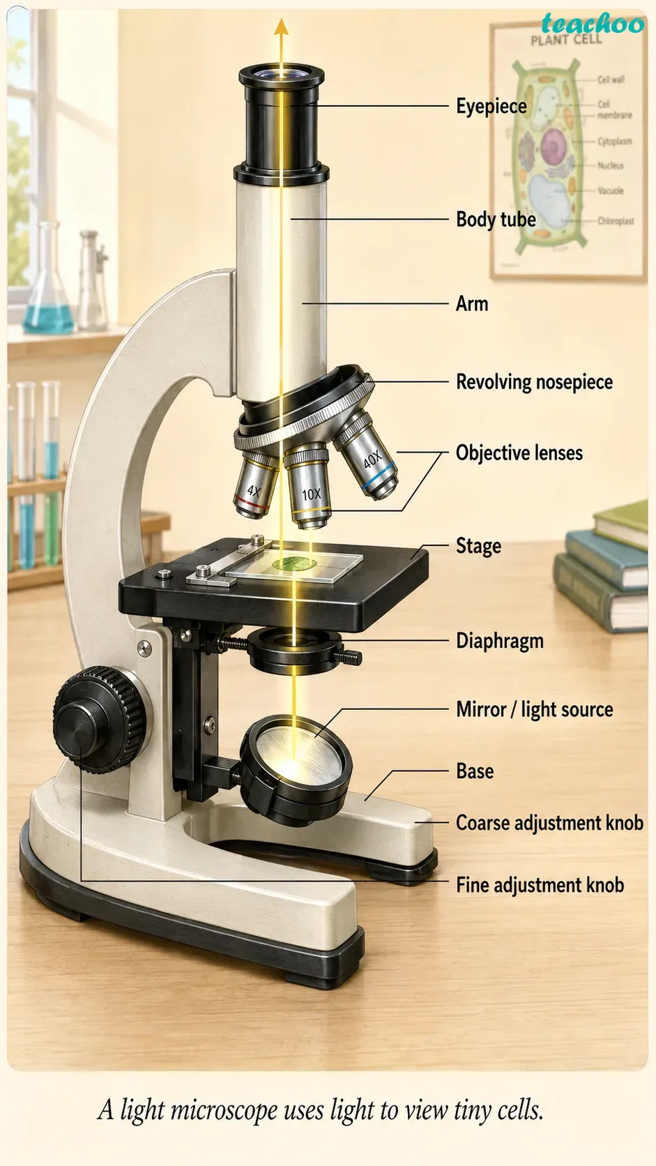
- It has an eyepiece, a body tube and objective lenses.
- Knobs (coarse and fine) focus the image.
- A stage holds the slide; a mirror sends in light.
In this Activity, we will measure the field of view of a microscope and use it to estimate the real size of one onion peel cell.
Activity 2.1 — Steps
Step 1
Place a mm ruler on the stage; measure the field-of-view diameter.
↓
Step 2
Convert the diameter from mm to micrometre (x1000).
↓
Step 3
Replace the ruler with an onion peel slide.
↓
Step 4
Count the cells across the diameter in a straight line.
↓
Cell size = field diameter / number of cells
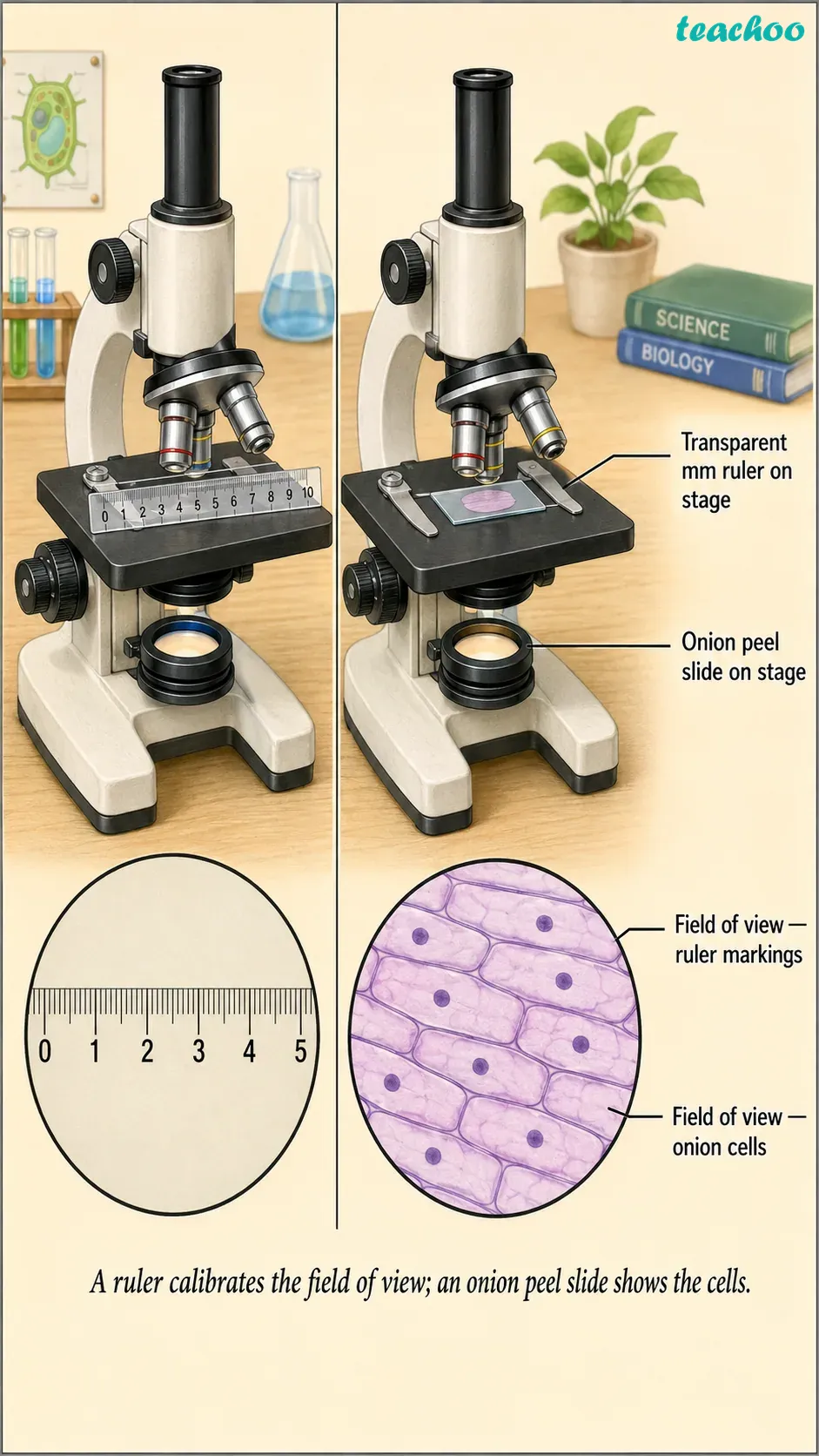
Materials needed
A microscope, a transparent ruler with mm markings, and an onion peel slide.
Procedure
1. Take a transparent ruler with millimetre (mm) markings.
2. Place the ruler on the stage, focus, and observe the diameter of the circular field of view through the eyepiece; measure it in mm.
3. Convert the diameter from mm to micrometre (µm). Suppose it is 5 mm, so 5 × 1000 = 5000 µm.
4. Remove the ruler and place an onion peel slide on the stage.
5. Focus on the slide and count the number of cells along the diameter in one straight line.
6. Estimate the real size of the cell using the formula.
2. Place the ruler on the stage, focus, and observe the diameter of the circular field of view through the eyepiece; measure it in mm.
3. Convert the diameter from mm to micrometre (µm). Suppose it is 5 mm, so 5 × 1000 = 5000 µm.
4. Remove the ruler and place an onion peel slide on the stage.
5. Focus on the slide and count the number of cells along the diameter in one straight line.
6. Estimate the real size of the cell using the formula.
Observation
Estimated size of onion peel cell = Diameter of the field in µm ÷ Number of cells along the diameter. If 25 cells are seen, size = 5000 µm ÷ 25 = 200 µm.
Explanation
A microscope makes a 200 µm cell appear larger. If both eyepiece and objective are 10X, the total magnification is 100X, so the cell looks 100 times bigger and clear.
◆ Summary
- Measure field width.
- Count cells across.
- Divide to get size.
- Microscope magnifies it.
What is an electron microscope?
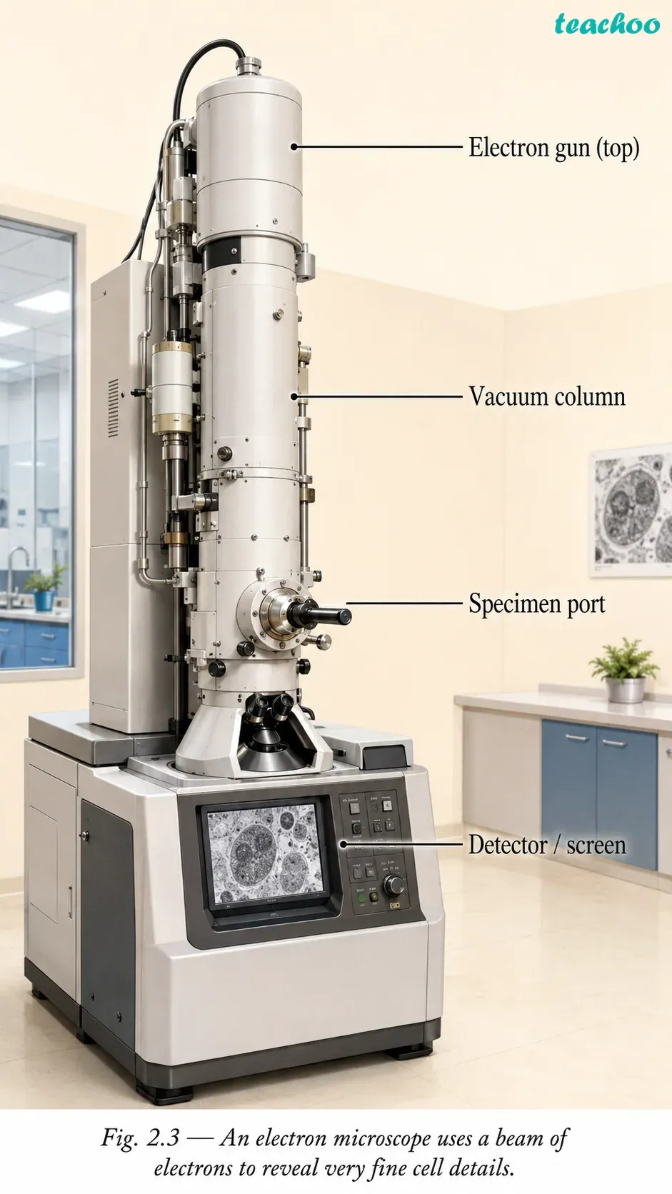
- It is a very powerful microscope.
- It uses a beam of electrons, not light.
- It shows fine cell structures at the nanometre scale.
- A nanometre is one-billionth of a metre.
Light microscope
Electron microscope
Uses light (visible).
Uses a beam of electrons.
Used in school labs.
Used by scientists for fine detail.
Shows cells and large parts.
Shows tiny parts at nanometre scale.
Important Points
- The limit of resolution of the human eye is 0.1 mm.
- Magnification = magnifying power of eyepiece × objective lens.
- Scientists improved microscopes by improving resolution, contrast and magnification.
💡 Ready to Go Beyond
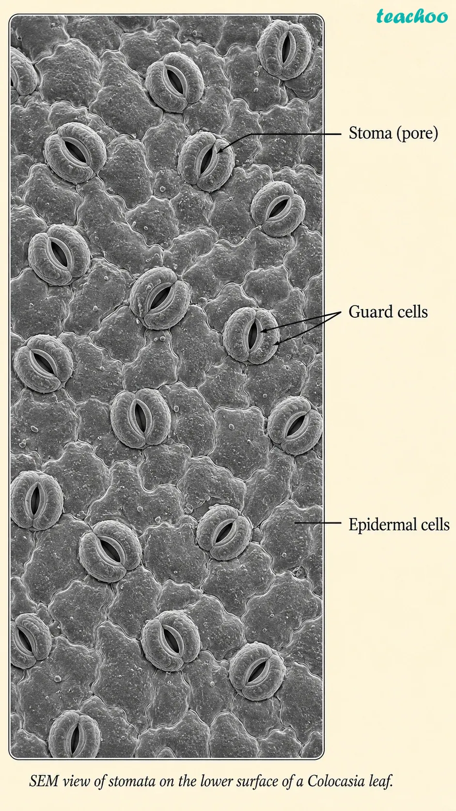
- Electron microscopes use a beam of electrons, not light.
- They reveal very fine details of a cell.
- They can show structures at the nanometre scale.
- A nanometre is one-billionth of a metre.
❓ Test Yourself
-
What is the limit of resolution of the human eye?
View Answer
About 0.1 mm. Two points closer than this look like one. -
Who first observed and named cells?
View Answer
Robert Hooke, in 1665, while looking at a slice of cork. -
What two lenses give a microscope its magnification?
View Answer
The objective lens and the eyepiece. -
If both lenses are 10X, what is the total magnification?
View Answer
100X. The cell appears 100 times larger. -
Which microscope uses electrons instead of light?
View Answer
The electron microscope. It shows the finest details.
Important Definitions
- Limit of resolution — the smallest distance at which two points are seen as separate (0.1 mm for the eye).
- Magnification — making a small object appear larger using lenses.
- Light microscope — a tool that uses light and lenses to view small objects.
- Electron microscope — a tool that uses a beam of electrons to see very fine details.